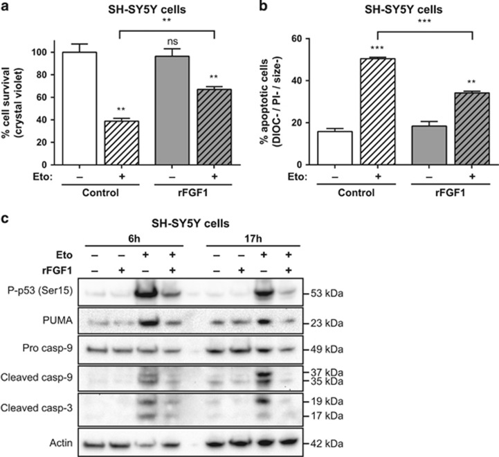
Figure 1.
Extracellular FGF1 protects SH-SY5Y cells from p53-dependent apoptosis. (a) SH-SY5Y cells were pretreated or not by adding recombinant FGF1 and heparin for 48 h (rFGF1) in the culture medium, then cells were treated or not with etoposide for 24 h (Eto). Cell survival was analyzed by crystal violet nuclei staining. (b) Following the same treatments, SH-SY5Y apoptotic cells were characterized by flow cytometry after DiOC6(3) and PI staining. Apoptotic cells correspond to the low DiOC6(3) (low ΔΨm, noted DIOC−) and low PI (to exclude necrotic cells, noted PI−) staining and small-sized cells (a hallmark of apoptotic cell condensation, noted size−). For (a and b), the graphs represent the mean±S.E.M. of three independent experiments. Student’s t-tests were performed relative to the control cells, except where indicated (n=3; n.s.: P>0.05; **P⩽0.01; ***P⩽ 0.001). (c) SH-SY5Y cells were pretreated or not with recombinant FGF1 (rFGF1) for 48 h, and then treated or not with etoposide (Eto) for 6 h or 17 h. Twenty micrograms of the corresponding cell lysate proteins were used to analyze by western blot the levels of P-p53 (Ser15) that reveals p53 activation, of the p53 proapoptotic target PUMA, of pro- and cleaved caspase-9 forms and cleaved caspase-3. Actin detection was used as a control

