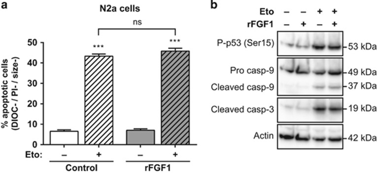
Figure 2.
Extracellular FGF1 does not protect N2a cells from p53-dependent apoptosis. (a) N2a cells were pretreated or not by adding recombinant FGF1 and heparin in the culture medium (rFGF1) for 48 h, then treated or not with etoposide (Eto) for 24 h. N2a apoptotic cells were characterized by flow cytometry after DiOC6(3) and PI staining (apoptotic cells are the DIOC−, PI− and size− cells). The graph represents the mean±S.E.M. of three independent experiments. Student’s t-tests were performed relative to control cells, except where indicated (n=3; n.s.: P>0.05; ***: P⩽0.001). (b) N2a cells were pretreated or not with recombinant FGF1 (rFGF1) for 48 h, and then treated or not with etoposide (Eto) for 24 h. Twenty micrograms of the corresponding cell lysate proteins were used to analyze by western blot the levels of P-p53 (Ser15) that reveals p53 activation, of pro- and cleaved caspase-9 forms and cleaved caspase-3. Actin detection was used as a control

