Figure 7.
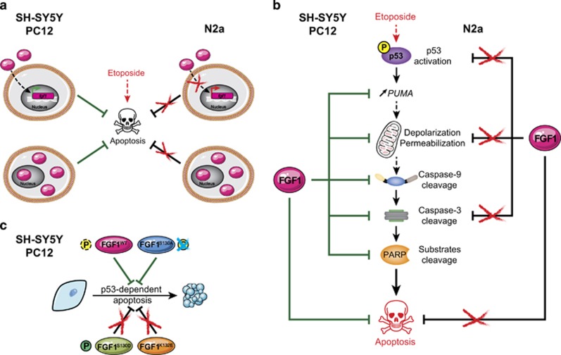
Role of extra- and intracellular FGF1 in neuronal SH-SY5Y, N2a and PC12 cells. (a) Both extra- and intra-cellular FGF1 protect human SH-SY5Y and rat PC12 cells from p53-dependent apoptosis (left). Endogenous fgf1 expression is increased by rFGF1 addition (top left). In contrast, extra- and intra-cellular FGF1 show no anti-apoptotic activity, and rFGF1 addition does not increase endogenous fgf1 expression in murine N2a cells (right). (b) Overexpression of intracellular FGF1 inhibits etoposide-induced apoptosis in human SH-SY5Y and rat PC12 cells by decreasing PUMA transactivation, mitochondrial membrane depolarization and permeabilization, and activation of caspase-9, caspase-3 as assessed by the cleavage of its substrate PARP (left). On the contrary, FGF1 overexpression does not protect N2a cells from etoposide-induced apoptosis, and modifies neither p53 activation, mitochondrial depolarization and permeabilization, nor cleavage of caspase-3 (right). (c) In human SH-SY5Y and rat PC12 cells, overexpression of wild-type FGF1 or non-phosphorylable FGF1S130A inhibits p53-dependent apoptosis, while overexpression of the phosphomimetic FGF1S130D or the FGF1K132E mutant does not. Therefore, phosphorylation of FGF1 inhibits its anti-apoptotic activity in SH-SY5Y and PC12 cells. This figure compiles the results of the present study performed in neuroblastoma SH-SY5Y and N2a cells and of previous studies performed in pheochromocytoma PC12 cells14, 15, 32

