Figure 4.
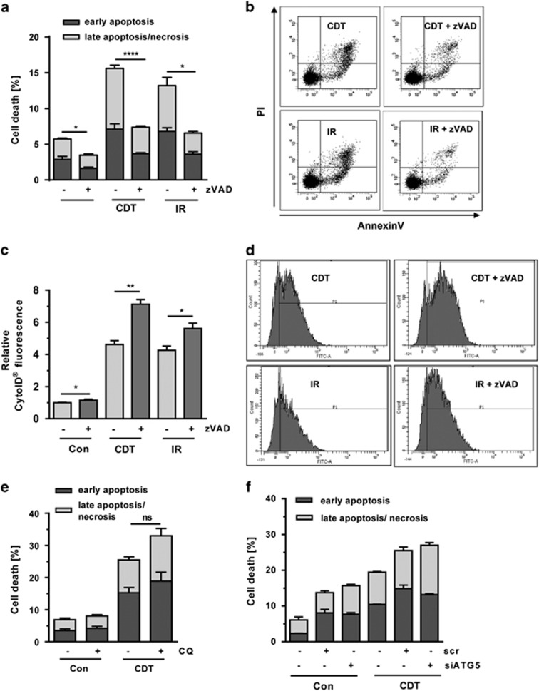
Interplay between DSB-induced autophagy and cell death. (a) Cell death induction by CDT and IR and involvement of caspases. HCT116 cells were treated with CDT (500 ng/ml) or exposed to IR (10 Gy). After 20 h, cells were supplemented with the pan-caspase inhibitor zVAD (20 μM) and incubated for another 28 h. Cell death was finally assessed by Annexin V/PI staining and flow cytometry (n=3); ***P<0.001, *P<0.05. (b) Representative dot plots of Annexin V/PI measurements. (c) Impact of pan-caspase inhibition on DSB-induced autophagy. HT116 cells were treated as described above and processed for CytoID staining followed by flow cytometry (n=3); **P<0.01, *P<0.05. (d) Representative histograms of CytoID staining. (e) Chemical inhibition of autophagy and DSB-induced cell death. HCT116 cells were exposed to CDT as described above and supplemented with CQ (10 μM). After 48 h, cells were harvested, stained with Annexin V/PI and analyzed by flow cytometry. (n=3); NS: not significant. (f) Impact of ATG5 knockdown on CDT-induced cell death. HCT116 cells were transfected with ATG5 siRNA or scrambled RNA. After 24 h, cells were exposed to CDT (500 ng/ml) and incubated for additional 48 h. Cell death induction was monitored by flow cytometry (n=3)

