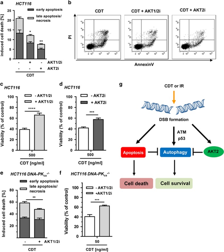
Figure 7.
Impact of AKT on cell survival following DSB induction. (a) Role of AKT and autophagy in DSB-induced cell death. Cells were exposed to CDT (500 ng/ml) for 48 h with or without AKT1/2 inhibition (1 μM) or AKT2 inhibition (1 μM). Cells were harvested and stained with Annexin V/PI. Early apoptotic and late apoptotic/necrotic cells were then determined by flow cytometry. (n=3); **P<0.01, *P<0.05. (b) Representative dot plots of Annexin V/PI staining. (c and d) Influence of AKT on cell viability upon DSB generation. Cells were incubated with increasing doses of CDT for 72 h in the absence or presence of AKT1/2 (0.1 μM) or AKT2 (5 μM) inhibitor. Viability was then assessed using MTS assay. (triplicates, n=3); ****P<0.0001, ***P<0.001, (e) AKT inhibition and DSB-induced cell death in DNA-PK-deficient cells. Cells were treated and processed as described under panel (a). (n=3); **P<0.01. (f) Impact of AKT on viability of DNA-PK-deficient cells upon DSB induction. Cells were challenged and viability was measured as mentioned in panel (b). (triplicates, n=3); ***P<0.001. (g) Proposed model of DSB-triggered autophagy and its regulation by AKT. IR or CDT-mediated DSB formation stimulates the autophagic flux in an ATM- and p53-dependent manner. Concomitantly, DSB formation provokes caspase-dependent apoptotic cell death, which suppresses autophagy. DSB-triggered AKT signaling, mainly attributable to AKT2, curtails pro-survival autophagy independent of DNA-PK. Inhibition of AKT signaling boosts autophagy and thereby cell survival

