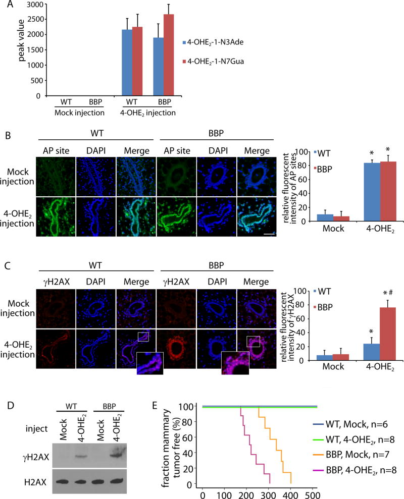
Figure 3. 4-OHE2 injection induces DNA lesions and breast tumor in vivo.
(A) 4-OHE2 injection induces 4-OHE2-1-N3Ade and 4-OHE2-1-N7Gua in mammary gland. Peak values of 4-OHE2-1-N3Ade and 4-OHE2-1-N7Gua from mammary gland of parous WT and BBP mice (10 weeks) in mass spectrometry are shown. (B, C) 4-OHE2 injection induces AP sites (B) and DSBs (C) in mammary gland of WT and BBP parous mice (14 weeks). Scale bars: 50 µm. Fluorescent intensity of AP sites (B) and γH2AX (C) was analyzed by Image J software. * and #, p < 0.01, respectively. (D) Phosphorylation of H2AX in mammary tissues from the injected WT and BBP mice was detected by Western blot. Total H2AX was used as the loading control. (E) Kaplan-Meier analysis shows the mammary tumor free fraction of WT and BBP mice injected with mock or 4-OHE2. The medians of tumor free BBP mice injected with mock or 4-OHE2 are 335 and 214 day, respectively.

