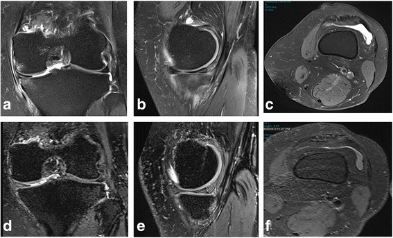
Fig. 1.

Pre-treatment 3T proton density fat saturated images in coronal (a) and sagittal (b) planes demonstrating medial compartment bone edema, more pronounced on the tibial side. Axial (c) proton density fat saturated imaging at the level of suprapatellar pouch demonstrating a knee effusion. Corresponding post-treatment 3T proton density fat saturated images in coronal (d) and sagittal (e) planes demonstrating resolution of medial compartment bone edema. Axial (f) proton density fat saturated imaging at the level of suprapatellar pouch demonstrating reduction in size of knee effusion
