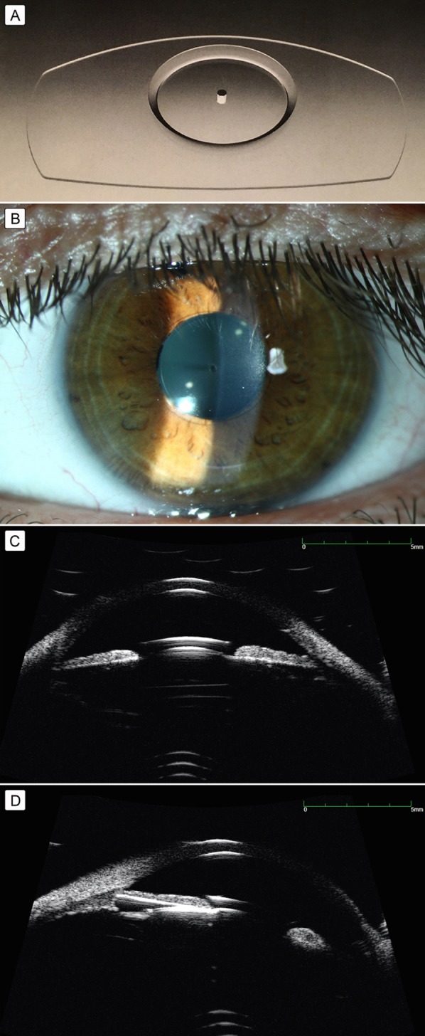
Figure 1.

A, Posterior chamber collamer phakic intraocular lens RSK-3 (S. Fyodorov Eye Microsurgery, Moscow, Russia). B, Slit-lamp view of the anterior segment. C–D, Anterior segment ultrasound biomicroscopy of the right eye showing collar-button-type collamer posterior chamber lens (C) and visualization of lens haptics (D).
