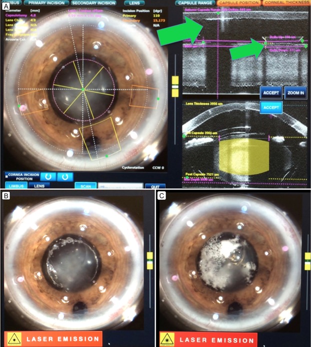
Figure 2.
Intraoperative femtosecond laser–assisted step view. A, Intraoperative view and optical coherence tomography (OCT) images of the anterior segment of the right eye showing blockage due to accumulation of emulsified silicone oil posterior to the corneal endothelium and PC pIOL optics margin. B, Intraoperative view of incomplete capsulotomy during the femtosecond laser step. C, Intraoperative view of trapped cavitation bubbles under the posterior chamber phakic intraocular lens.

