
Figure 3.
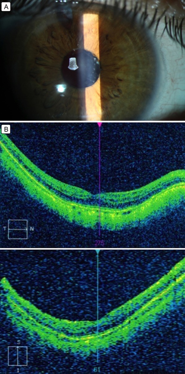
Postoperative diagnostic data. A, Slit-lamp view of the anterior segment on postoperative day 1. B, Optical coherence tomography of the macular area; retinal layers attached. There is sectoral thinning of the superior paracentral, inferior paracentral and peripheral retinal nerve fiber layers in the macular area.