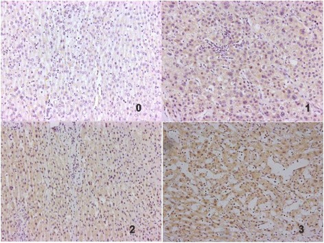
Fig. 5.

Immunohistochemical staining of INTS6 in HCC. INTS6 protein expression localized mainly to the nuclei in tumour cells. Different INTS6 staining intensities [negative: 0, weak: 1, moderate: 2, strong: 3] are indicated in the micrographs

Immunohistochemical staining of INTS6 in HCC. INTS6 protein expression localized mainly to the nuclei in tumour cells. Different INTS6 staining intensities [negative: 0, weak: 1, moderate: 2, strong: 3] are indicated in the micrographs