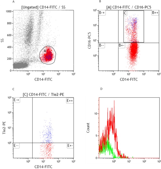
Fig 1. Identification of TEMs as CD14+CD16+TIE2+ cells in peripheral blood.

A. CD14+ PBMC obtained from HCC patients were stained and analyzed by flow cytometry. B. CD14+ monocytes were divided into two distinct subsets, CD14+CD16+ and CD14+CD16- cells. C. TEMs were examined by using TIE2-PE orIgG1-k-PEisotypic antibody. D.The percentage of TEMs in peripheral blood CD14+CD16+ monocytes for this sample was 16.14% (the percentage of TEMs in CD14+CD16+ monocytes measured by TIE2-PE staining minus that measured by IgG1-k-PE staining, 24.2% - 8.06% = 16.14%).
