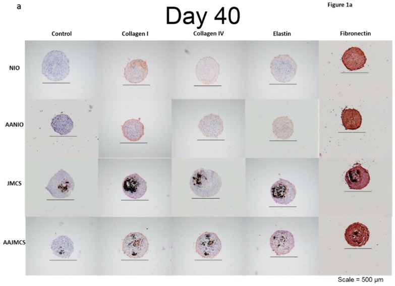
Figure 1.
Immunohistochemistry staining of spheroids over time. Spheroids containing iron oxide MNPs and spheroids without MNPs were immunohistochemically examined for collagen I, collagen IV, elastin, and fibronectin after a 40-day incubation period in cell culture media. Spheroids with and without MNPs were also incubated in media supplemented with ascorbic acid, which is a known stimulant of collagen production. Each spheroid type demonstrated positive stains for each ECM marker when compared to their respective controls. A preferential cellular organization, as illustrated in the spherical geometry of the spheroids was observed (black = iron oxide, brown = positive stain).

