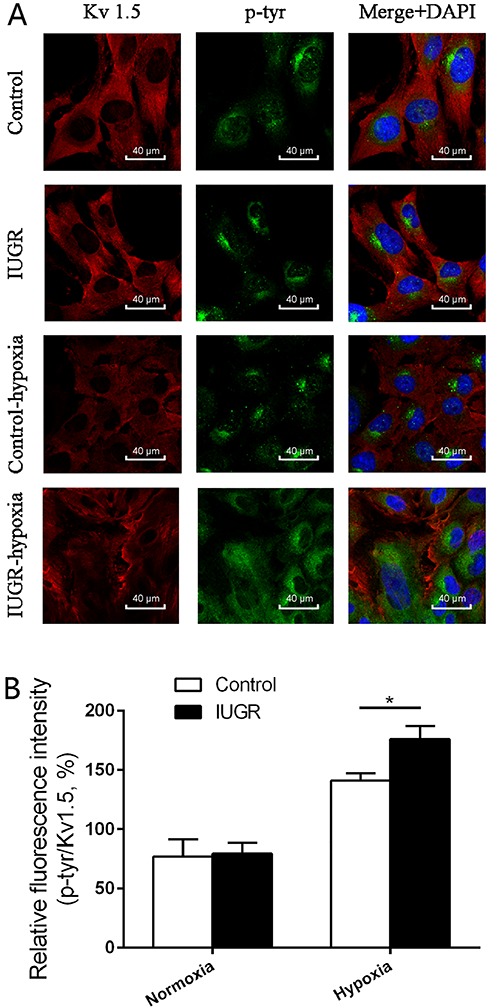
Figure 4. Immunofluorescence detection of Kv1.5 and tyrosine-phosphorylated Kv1.5 in pulmonary artery smooth muscle cells PASMCs. A, Kv1.5 expression (red) is downregulated and tyrosine-phosphorylated Kv1.5 expression (green) is upregulated to varying degrees in both intrauterine growth retardation (IUGR) rats and control rats after 2 weeks of hypoxia. Nuclei are stained with DAPI. Scale bar=20 µm. B, Summary histogram of relative fluorescence intensity of p-tyr/Kv1.5. Data are reported as means±SD. *P<0.05 (ANOVA).