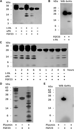
Fig. 4. Proteolysis of rhFGF23 by tPA, uPA, and plasmin in vitro.

(A) Silver-stained SDS-PAGE gel of reactions containing purified recombinant human tPA, uPA, and FGF23. (B) Western blot (WB) using anti–6×His-tag antibody and tPA reactions from (A). (C) Silver-stained SDS-PAGE gel of in vitro time courses of FGF23 proteolysis by tPA and uPA. (D) Silver-stained gel showing degradation of FGF23 by plasmin. (E) Immunoblot of the gel in (D) with anti–6×His-tag antibody shows iFGF23 (lane 2) and a complete degradation of FGF23 by plasmin (lane 3).
