
FIGURE 8:
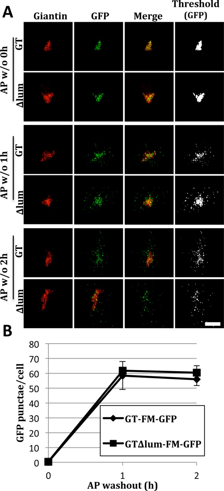
Lysosomal redistribution of GT∆lum-FM-GFP. (A) Cells were transfected with either GT-FM-GFP or GTΔlum-FM-GFP (all GT lumenal sequence removed) and subjected to an AP washout for 0, 1, or 2 h before staining for giantin and visualization of GFP. Merged and thresholded (GFP only) images are also shown. (B) Quantification shows the number of GFP punctae for the constructs at each time point (mean ± SEM, n = 3, >12 cells per experiment, p ≥ 0.3).