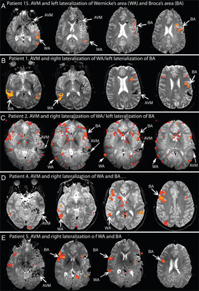
Fig. 4.
Example fMR imaging language activations. A: Case 15 (Group 2) exhibits left dominance of both the Broca area (BA) and Wernicke area (WA). B and C: Cases 1 and 2 (Group 1) demonstrate right dominance of Wernicke area and left dominance of Broca area. D: Case 4 (Group 1) has an AVM that potentially disrupts the arcuate fasciculus. There is right dominance in both Wernicke area and Broca areas. E: Case 5 (Group 1) has a lesion that also likely disrupts the arcuate fasciculus. The patient exhibits right dominance of both Wernicke area and Broca areas.

