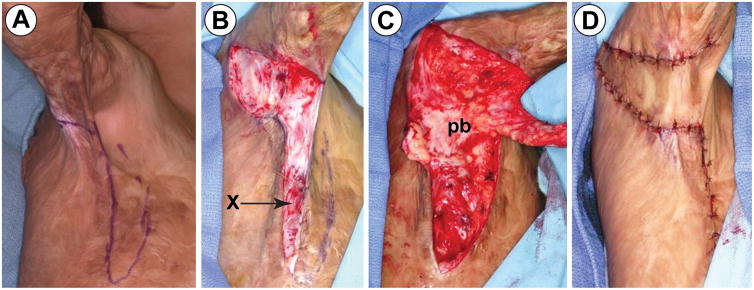
Figure 1.
Transpositional random fasciocutaneous flap technique: (A) Right triangle of skin showing the cathetus adjacent, which is perpendicular to the line of release. (B) An incision is made along the cathetus adjacent following the release of contracted area. The incision is continued down to the fascial layer and the fascia is anchored to the skin edge. (“x” mark). Identification of the vascular pedicle is not necessary. (C) A random fasciocutaneous flap is fabricated. Blunt dissection is used to mobilize the pedicle to minimize damage to the vascular pedicle. (D) The random fasciocutaneous flap is rotated 90° for flap inset. All wound edges are closed primarily.

