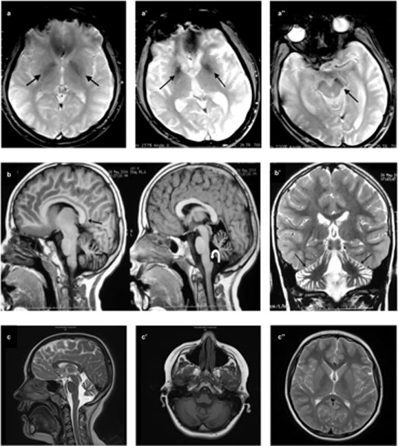
Fig 2.

MRI findings. Axial gradient-recalled echo image shows bilateral hypointensity in both globi pallidi (arrows) consistent with iron accumulation in Case 1 at age 27. (a). Axial gradient-recalled echo sequence of Case 2 at age 32 depicts hypointensity in both globi pallidi (a′) and substantia nigra (a″) (arrows) consistent with iron accumulation. Sagittal T1-weighted images demonstrate callosal elongation (black arrow) and claval hypertrophy (curved white arrow) (b), marked atrophy of cerebellum on T2-weighted image (b′) of Case 3 at the age of seven. T2-weighted sagittal scan shows vermian atrophy and claval hypertrophy (arrow) (c), T1-weighted axial image demonstrates cerebellar atrophy (c′), and there was no evidence of iron deposition in basal ganglia on T2-weighted axial scan (c″) in Case 4 at age 10.
