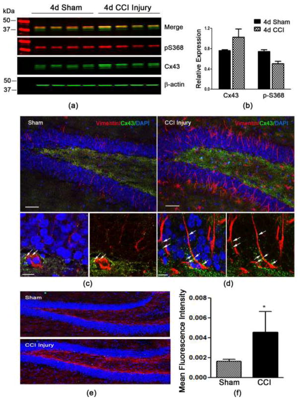
Fig. 1. Cx43 expression in the hippocampus 4 days post-CCI injury.
(a) Western blot analysis showing total Cx43 and phospho (p) serine 368 protein levels in the whole hippocampus at 4 days post-sham or CCI injury. (b) Quantified data showing the fluorescence intensity of Cx43 relative to β-actin and p-S368 relative to total Cx43. (c) Immunohistochemistry for vimentin (red) and Cx43 (green) expression in the dentate gyrus (DG) of sham injury mice at 4 days post-sham compared to CCI injury (d). Increased expression by immunofluorescence is seen in the DG following CCI injury (e). (f) The mean fluorescence intensity of Cx43 expression in the region of the subgranular layer of the DG was significantly increased after CCI injury. *P<0.01 compared to sham injury.

