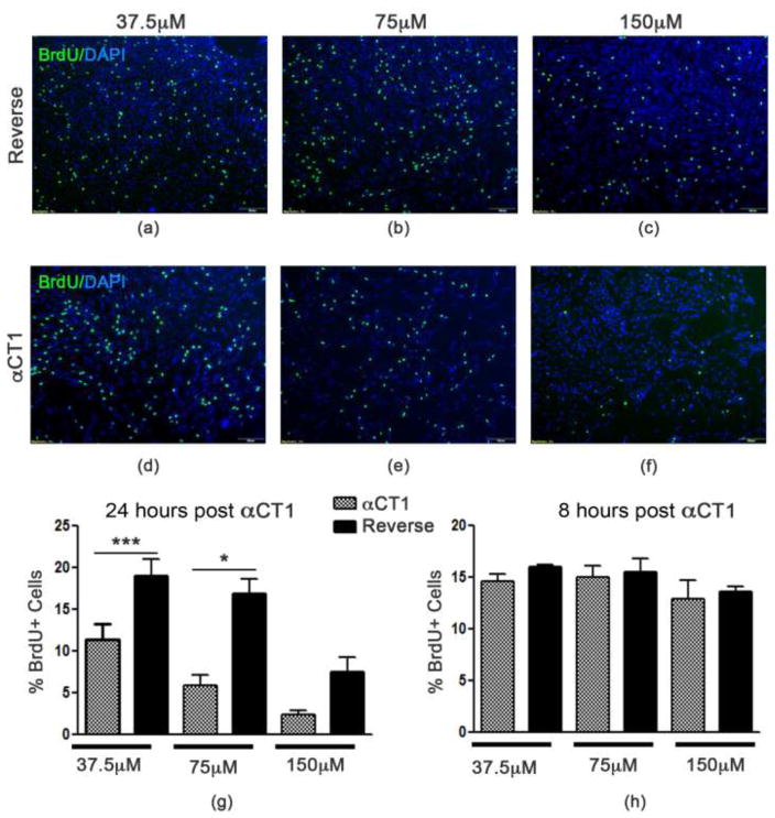
Fig. 2. Proliferation of NSPCs is reduced following αCT1treatment.
Exposure of NSPCs to selective inhibitor αCT1 (d–f) reduces BrdU incorporation in a dose dependent manner following 24 hour incubation compared to the control Reverse control peptide (a–c). (g) Quantified data showing a significant reduction in the percentage of BrdU-positive cells at 37.5 and 75 μm αCT1 exposure compared to Reverse. Non-specific effects of Reverse at 150 μm were also seen. (h) Quantified data showing no changes in NSPC proliferation are evident at 8 hours of αCT1 treatment. *P<0.01; ***P<0.001 compared to Reverse control peptide.

