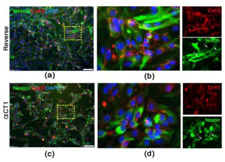
Fig. 4. αCT1 reduces Cx43 expression and induces changes in NSPC morphology.

(a–b) Immunocytochemistry using antibodies against Cx43 (red) and Nestin (green) on monolayer cultures of NSPCS treated with 150 μm Reverse control peptide for 24 hours. (c–d) Significant morphological changes are seen following 150 μm αCT1 exposure, demonstrated by nestin staining showing NSPCs are more rounded with fewer protrusions. The density and aggregate number and size of Cx43 is also visibly reduced compared to Reverse control peptide.
