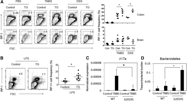
Figure 2. Increased myeloid cell expansion in the intestine and blood, but not the brain, of LRRK2 G2019S rats challenged with TNBS, DSS, or LPS.
(A) RP-1+ cell frequency was examined in the colonic lamina propria. Control and LRRK2 G2019S TG rats were administered TNBS (rectal injection, 50 mg/kg of body weight) or DSS (4∼5%) as described in Supplemental Fig. 1. Rats were sacrificed 6 (TNBS) or 25 (DSS) d later, and indicated organs and tissues were examined for RP-1+ cell frequency. (B) Control and LRRK2 G2019S TG rats were injected with LPS (1.2 million EU/kg per on d 0) and sacrificed 15 d later, and blood cells were examined for RP-1+ cell frequency. (C) Colon tissues were examined for il17a mRNA expression. Expression levels were normalized by that of β-actin. (D) Colon tissues were examined for levels of Bacteroidetes based on Bacteroidetes-specific 16S rRNA gene levels. Tissue Bacteroidetes levels were normalized by β-actin gene levels. Representative dot plots and pooled data (n = 3–6) from 2 individual experiments are shown. *Significant differences (P ≤ 0.05) from control groups. Error bars indicate means ± sem.

