. 2017 Aug 28;26(4):213–226. doi: 10.5607/en.2017.26.4.213

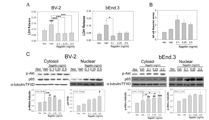
Fig. 9. Treatment of flagellin increased cell survival in hypoxic conditions. (A) After 2 h OGD, cell cytotoxicity levels were measured by a LDH assay at 1 h reperfusion. The flagellin-treated groups had lower cell cytotoxicity than the vehicle group. Up to 0.5 ng/ml, flagellin had protective effects on BV-2 and bEnd.3 cells. Data are mean±SE. n =4 (###p<0.001 compared with normoxia group; *p<0.05 and ***p<0.001 compared with the vehicle group). (B) NF-κB activity was detected by a reporter gene assay in bEnd.3 cells 1 h after reperfusion. In the vehicle group, the expression of the NF-κB promoter was increased compared to the normoxia group. When cells were treated with flagellin, the activity of NF-κB was increased compared to the vehicle group. Data are mean±SE. n=3. (C) Western blot analysis revealed that flagellin treatment increased the phosphorylation of Akt compared to vehicle treatment after 1 h reperfusion. Flagellin treatment increased the nuclear translocation of p65 compared to vehicle at the same time point. Data are mean±SE. n=4 (#p<0.05, ##p<0.01, and ###p<0.001 compared with normoxia group; *p<0.05 and **p<0.01 compared with the vehicle group).

Fig. 9