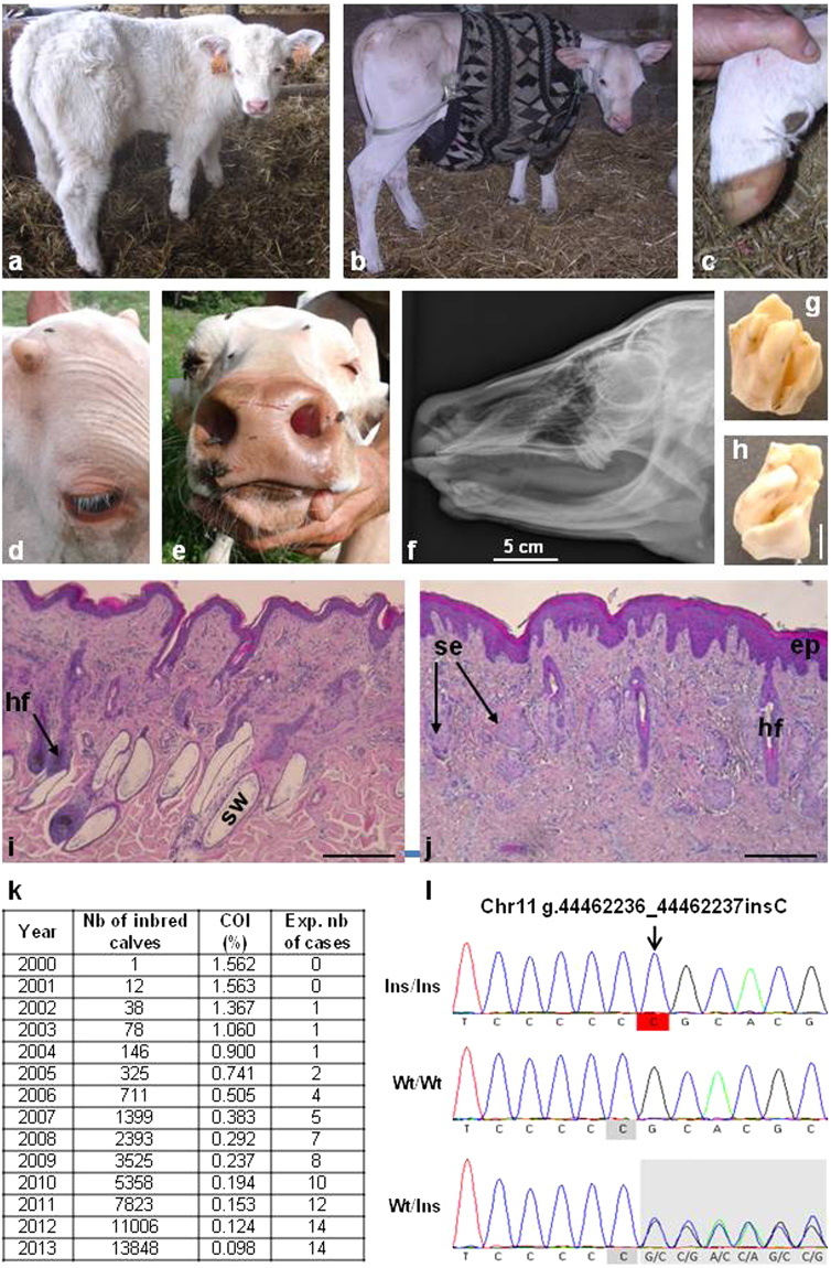
Figure 6.
Identification of a de novo mutation causing recessive anhidrotic ectodermal dysplasia (AED) before the emergence of this defect in the population. (a) One-month old control calf. (b) One-month old AED affected calf showing generalized hypotrichosis. (c) Hoof of the same AED calf with no notable malformations. Note that in humans most AED patients have normal nails too. (d) Detail of the head of a five-month-old AED calf, showing eyelashes and normal development of horns. (e) Muzzle of the same calf showing normal vibrissae and extreme skin dryness due to the absence of nasolabial glands (see also Supplementary Fig. 19). (f) X-ray of the skull of a one-month old calf showing a complete absence of incisors and the presence of only one upper molar on each side. (g,h) Detail of one upper molar showing abnormal cusps and deep grooves. (i,j) Histological sections of skin biopsies from control (i) and AED (j) one-month-old calves. In AED animals, the epidermis (ep) is acanthotic (i.e. thickened). The number of hair follicles is greatly reduced and deep portions of them are atrophic or absent. Sweat glands (sw) are absent. Sebaceous glands (se) appear normal but a number of them are “orphan” (i.e. without associated hair follicle). Scale bars represent 500 µm. (k) Yearly estimation of the number of calves predicted to be born homozygous for the g.44462236_44462237insC mutation on chromosome 11 which is predicted to cause a frameshift and premature truncation of EDAR. The total numbers of calves with both parents related to primo-mutant bull Invincible, as well as their coefficient of inbreeding (COI) are also presented. (l) Electrophoregrams of a homozygous (Ins/Ins) AED affected calf, a homozygous wild-type (Wt/Wt) animal and a heterozygous carrier of the mutation (Wt/Ins).

