Figure 1.
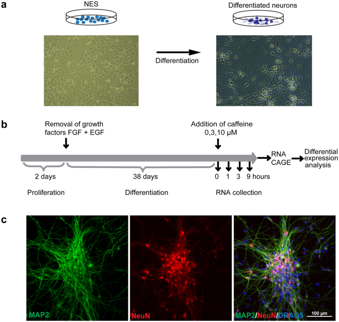
Experimental design. (a) NES cell differentiation: bright field images of NES cells and differentiated neurons. (b) Timeline of the experiment. NES cells were plated and kept in the presence of FGF and EGF for two days. Growth factors were removed and cells were differentiated for 38 days. Caffeine was added and RNA was collected after 0 h, 1 h, 3 h and 9 h. The samples were then used for CAGE sequencing and differential expression analysis. (c) Phenotype of differentiated cells. After 38 days, cells were fixed and stained for the neuronal markers MAP2 and NeuN and the nuclear marker DRAQ5. Untreated cells are shown.

