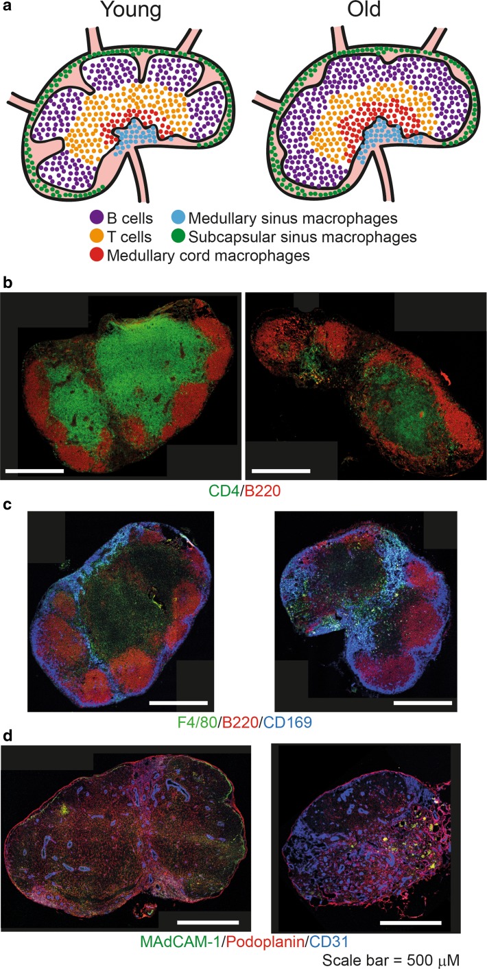
Fig. 3.
Structural organisation of lymph nodes and changes with age. a In a young mouse subcapsular sinus macrophages are located underneath the capsule. On the afferent side of the lymph nodes B cell follicles form distinct follicles in the cortex. Adjacent to the follicle are the T cells in the paracortical area. In the medullary region medullary cord and medullary sinus macrophages are found. In aged lymph nodes B cell follicles no longer form distinct follicles and there is an increase in macrophage populations, especially in the medullary region. b Immunofluorescent staining shows changes to lymph nodes with age. Aged lymph nodes no longer form distinct B cell follicles (B220+) and there are less T cells. c There are increased amounts of subcapsular sinus macrophages (CD169+, F4/80−), medullary sinus macrophages (CD169+, F4/80+) and medullary cord macrophages (CD169−, F4/80+) in aged lymph nodes. d Stromal cells are also disrupted with age. An increase in blood endothelial cells (CD31+) which form high endothelial venules is evident and are more widely spread throughout the lymph node. Images of inguinal lymph nodes from 2 and 18 month old C57BL/6 mice were acquired by confocal microscopy. Features of this figure are reproduced from (Turner and Mabbott 2017b) under the terms of the CC-BY-NC Creative Commons Attribution Licence 4.0 (Turner and Mabbott 2017b)

