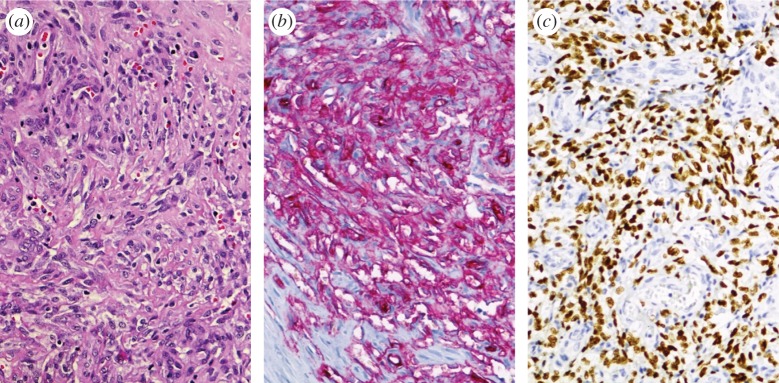
Figure 1.
Histology of a KS tumour infiltrating a lymph node (250× magnification). (a) HE staining showing the typical histological features such as elongated spindle cells, abnormal vessels with thinned epithelium and extravasated erythrocytes. (b) Immunohistochemistry staining for CD34 to indicate the endothelial origin of the spindle cells. (c) Immunohistochemistry staining for LANA, showing tumour cells with a latent KSHV infection.

