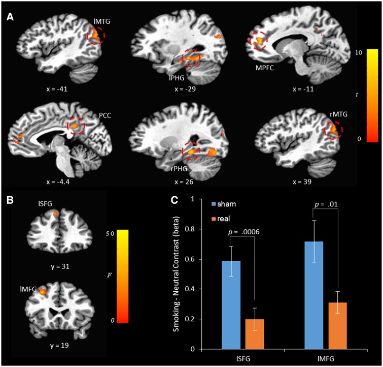
Fig. 2.
(A) The smoking-neutral contrast map in the cue-reactivity task. l: left; r: right; MPFC, medial prefrontal cortex; PHG, para-hippocampal gyrus; FG: right fusiform gyrus; MTG: middle temporal gyrus; PCC, posterior cingulate cortex; SFG: superior frontal gyrus; MFG: middle frontal gyrus. The left, the posterior, and the inferior directions are negative. The contrast map was thresholded with cluster-wise P values <0.05. (B) tDCS × cue type interaction. (C) A detailed illustration of the tDCS × cue interaction in the left SFG and the left MFG.

