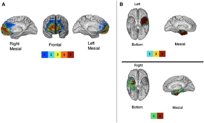
Fig. 1.
(A) Mesial and frontal views of the overlap map of lesions for the five vmPFC patients. The area of maximal overlap lies in the vmPFC. (B) Bottom and left hemisphere mesial views for of the overlap map of lesions for the three left amygdala patients and bottom and right hemisphere mesial views of the overlap map of lesions for the two right amygdala patients are presented. The highest overlap in the vmPFC group includes medial frontopolar areas and the highest overlap in the left amygdala group includes the temporal pole and perirhinal areas. For both Panel A and B, the color bar indicates the number of overlapping lesions at each voxel.

