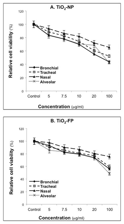
Figure 1. Viability of TiO2-exposed airway epithelia.

Human nasal, tracheal, bronchial and alveolar epithelial cells were exposed to various concentrations of TiO2-NP (A) or TiO2-FP (B) for 24 hr. Relative cell viability was measured by the MTT assay. Data are expressed as means ± SDs of 3 independent experiments.
