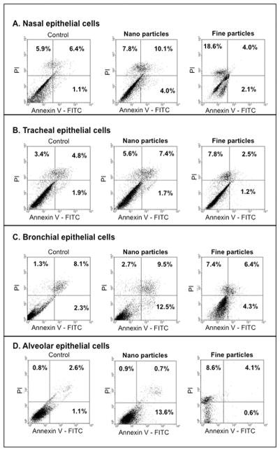
Figure 7. Nano vs. fine particles-induced cell death.
FACS data for nasal (A), tracheal (B), bronchial (C) and alveolar (D) epithelial cells are shown. Airway epithelial cells were exposed to 10 μg/ml of TiO2-NP or TiO2-FP, then stained with annexin V-FITC and propidium iodide (PI), and cytotoxicity was measured by FACS. Bronchial and alveolar epithelial cells showed increased fractions of early apoptotic cells after TiO2-NP exposure, while TiO2-FP diverted cells to necrosis. Data shown are representative of 3 independent experiments.

