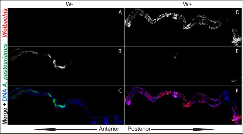
FIG 4 .

A. pasteurianus is absent from Wolbachia-infected (W+) L3 larval guts. (A to F) Composites of z-stack projections of confocal images of gnotobiotic L3 larval midguts. (A to C) Wolbachia-free (W−) guts. (D to F) Wolbachia-infected guts. (A, D) Wolbachia channel. (B, E) A. pasteurianus channel. (C, F) Merged images of Wolbachia (red), A. pasteurianus (green), and DNA (blue).
