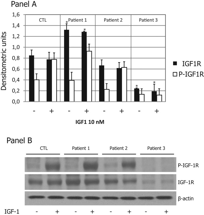
Figure 2.
IGF1R autophosphorylation and IGF1R protein expression. Lymphocytes isolated as described in the ‘Materials and methods’ section were lysed in RIPA buffer. Panel A: Cell lysates underwent to SDS-PAGE and the resolved proteins were transferred to nitrocellulose membranes, immunoblotted with anti-phospho-1R/IGF1R (Tyr1158/Tyr1162/Tyr1163) antibody and detected by ECL. The nitrocellulose membrane was then stripped with buffer Restore and, subsequently, reprobed with an anti-IGF1R rabbit polyclonal antibody. The membranes were blotted with an anti-β-actin antibody to control for protein loading. Panel B: Densitometric analysis was performed on two independent experiments. Data are the mean ± s.e.m. of two independent experiments. *P < 0.05. CTL, control; P-IGF1R, phosphorylated IGF1R.

 This work is licensed under a