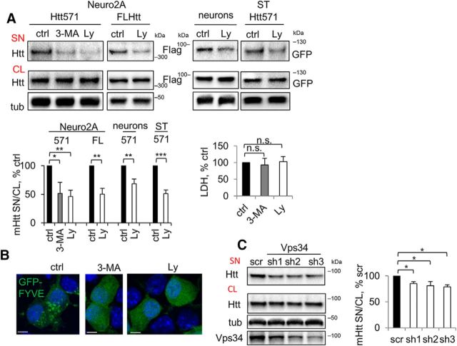
Figure 6.
Inhibition of PI3K reduces the secretion of mHtt. A, Reduced secretion of mHtt by inhibition of PI3K. Top, Neuro2A-mHtt cells were treated for 3 h with DMSO (ctrl), 10 mm 3-MA, or 25 μm Ly294002 (Ly); naive Neuro2A cells were transfected with FL Htt, primary cortical neurons were transduced with Htt571/72Q-Flag lentivirus and STHdh+/Hdh+ were transfected with Htt 571/72Q-GFP. Cells were then treated for 3 h with DMSO or Ly294002. Concentrated media and cell lysates were analyzed by Western blotting using anti-Htt, anti-Flag, or anti-GFP antibodies. Tubulin was used as a loading control. Bottom left, Ratio of mHtt in the media and cell lysates expressed as a percentage of control. n = 3, p = 0.0126, 0.0011, 0.0012, 0.0031, and 0.0003. Bottom right, The LDH assay was performed on 30 μl of the media from Neuro2A-mHtt cells before concentrating. n = 3, p = 0.4760 and 0.6717. B, 3-MA or Ly294002 treatment disrupts vesicular targeting of pEGFP-2xFYVE efficiently. Neuro2A-mHtt cells were transfected with pEGFP-2xFYVE. Twenty hours after transfection, the cells were treated for 3 h with DMSO (ctrl), 10 mm 3-MA, or 25 μm Ly294002 (Ly), fixed, and analyzed by confocal microscopy. Nuclei were visualized using DAPI staining. Scale bar, 10 μm. C, Knock down of Vps34 leads to reduced secretion of mHtt. Left, Cells were transfected with scrambled shRNA or shRNAs against Vps34 and incubated for 3–4 d. Media were replaced with OptiMEM 16 h before harvesting of the media and cell lysis. Concentrated media and the cell lysates were analyzed by Western blotting using anti-Htt antibody. Tubulin was used as a loading control. Knock down of Vps34 was verified by probing with anti-Vps34 antibody (bottom). Right, Ratio of mHtt in the media and cell lysates expressed as a percentage of control. n = 3, p = 0.0338, 0.0161, and 0.0170. Error bars indicate SD. *p < 0.05; **p < 0.01; ***p < 0.001; n.s., Not significant.

