Figure 7.
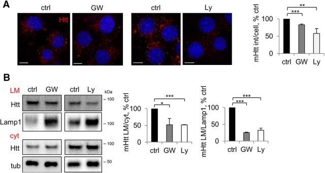
NS and PI3K inhibition impair vesicular targeting of mHtt. A, Punctate mHtt is reduced by inhibition of NS or PI3K. Neuro2A-mHtt cells were treated for 16 h with DMSO (ctrl) or 5 μm GW4869 (GW) (left) and for 3 h with DMSO or 25 μm Ly294002 (Ly) (right), followed by pre-permeabilization, fixation, immunolabeling with anti-Htt antibody, and analysis by confocal microscopy. DAPI was used to visualize nuclei. Scale bar, 10 μm. Right, Integrated density of mHtt signal per cell expressed as a percentage of control. n = 3, p = 0.0001 and 0.0067. B, mHtt in the light membrane fraction is reduced by inhibition of NS or PI3K. Left, Neuro2A-mHtt cells were treated for 16 h with DMSO (ctrl) or 5 μm GW4869 (GW) and for 3 h with DMSO (ctrl) or 25 μm Ly294002 (Ly). PNS were subjected to floatation in the sucrose density gradient and 2 μg of cytosolic (cyt) and light membrane (LM) protein equivalents were analyzed by Western blotting using anti-Htt antibody. Lamp1 and tubulin were used as LM and cyt markers, respectively. Center, Ratios of mHtt in the LM and cyt expressed as a percentage of control. n = 3, p = 0.0111 and 0.0001. Right, Ratios of mHtt and Lamp1 in the LM expressed as a percentage of control. n = 3, p = 0.0001 and 0.0001. Error bars indicate SD. *p < 0.05; **p < 0.01; ***p < 0.001.

