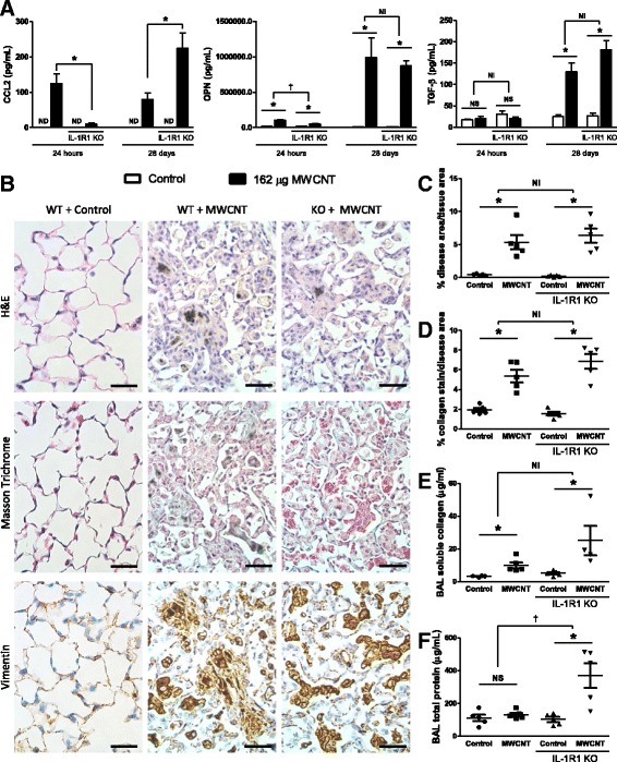
Fig. 3.

Fibrotic disease is not attenuated 28 days after MWCNT exposure. C57BL/6 and IL-1R1 KO mice were intratracheally administered 162μg of MWCNTs, and samples were collected 24 h and 28 days later. a The pro-fibrotic genes CCL2, osteopontin (OPN), and total TGF-β levels including the active form were measured by ELISA. b Representative histology images of the diseased area of lungs 28 days after MWCNT instillation compare WT and IL-1R1 KO mice. These images were obtained from slides stained with H&E, Masson Trichrome for collagen deposition (blue), and immune-staining for Vimentin, a surface marker of fibroblasts (brown). The scale bar represents 50 μm. c Entire longitudinal cross sections of the lungs were imaged and the disease area verses the total lung area were determined to quantify the pathology in H&E stained samples 28 days post exposure. d Representative images of pathology were taken of the Masson trichrome stained slides and the amount of collagen positive stain was quantified and normalized as a percent of area imaged. Non-diseased areas from exposed mice were used as controls. e Soluble collagen was measured in the BAL fluid at the 28 day time point, and f total protein was measured in BAL as well. Data represent mean ± SEM. n = 4–5. Statistical analysis was performed using two-way ANOVA. *p < 0.05, NS = not significant, †statistical interaction with p < 0.05, and NI = no statistical interaction
