
Fig. 1.
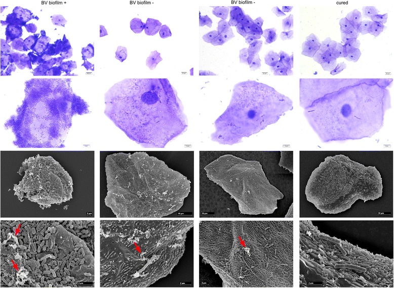
Microscopic images of vaginal epithelial cells with attached biofilms. From left to right: Vaginal epithelial cells from patients with BV either with (BV biofilm +) or without biofilm (BV biofilm −) and after metronidazole treatment (cured). Top two rows: light microscopy of samples stained with crystal violet. Bottom two rows: scanning electron micrographs. Red arrows indicate EPS.