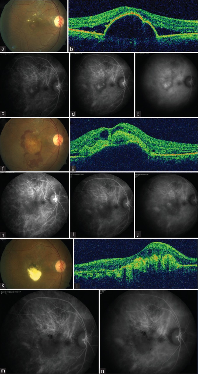
Figure 1.

At baseline, color fundus photograph of 63-year-old male reveals serosanguineous detachment of the posterior pole (a), with a peaked pigment epithelium detachment on optical coherence tomography (b), and indocyanine green angiography revealing clusters of polyps (c-e). Best-corrected visual acuity was 0.5 logarithm of the minimum angle of resolution. The patient underwent reduced-fluence photodynamic therapy. Five weeks later, the patient presented with hemorrhage (f), optical coherence tomography revealed hemorrhagic pigment epithelium detachment and cystoid retinal changes (g), and regressing polyps on indocyanine green angiography (h-j). Best-corrected visual acuity was 0.5 logarithm of the minimum angle of resolution. Two months later, subretinal fluid resolved with subfoveal aggregation of exudates (k and l), and lack of polypoidal activity on indocyanine green angiography (m and n). Best-corrected visual acuity was 1.7 logarithm of the minimum angle of resolution. The patient was periodically observed without the need for further treatment and remained stable up to the last follow-up at 53 months. Best-corrected visual acuity was 1.3 logarithm of the minimum angle of resolution
