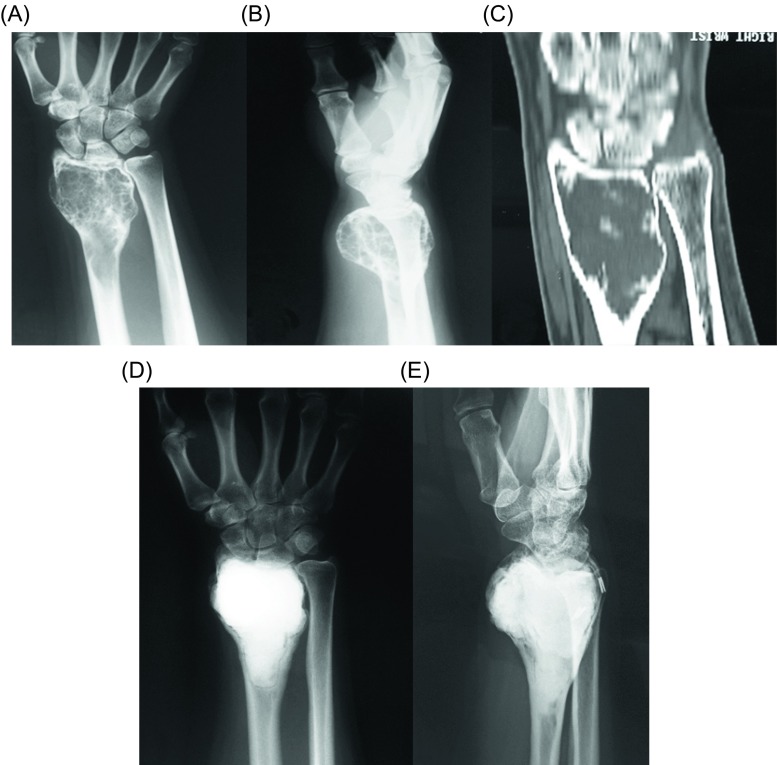
Figure 5.
(A) Anteroposterior and (B) lateral radiographs, and (C) coronal CT of the right wrist of a 43-year-old man with a GCT of the distal radius. Curettage, cauterization, and PMMA cementation were done. (D) Anteroposterior and (E) lateral radiographs of the right wrist show no evidence of local tumor recurrence at eight-year follow-up.

