Figure 4.
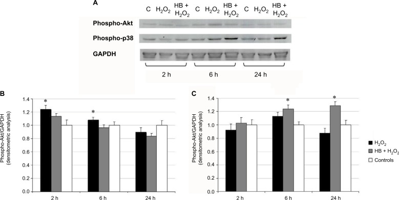
HB induced p38 phosphorylation and slightly reduced Akt activation.
Notes: (A) Western blot analysis on total cell lysates after treatment with HB and H2O2 and H2O2 alone. GAPDH was considered as a loading marker. Control was represented by untreated cells (not treated with HB or H2O2). (B) Densitometric analysis of phospho-Akt and GAPDH bands indicated an increased phosphorylation of Akt after oxidative stress induction by H2O2, but this effect disappeared in HB-pretreated cells. (C) Densitometric analysis of phospho-p38 and GAPDH indicated that HB pretreatment induced p38 activation. Data are given as mean ± SEM of three independent experiments. Statistical analysis: one-way ANOVA (n = 3). Significantly different from H2O2-treated cells (*p < 0.05).
Abbreviations: HB, hyaluronic acid and butyric acid; SEM, standard error of the mean; ANOVA, analysis of variance.

