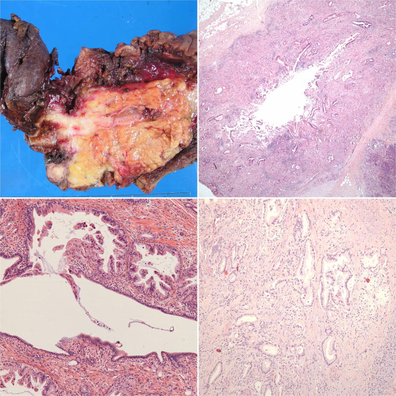
Figure 3.
(a) This pancreatoduodenectomy specimen demonstrates a distal common bile duct carcinoma forming a constrictive lesion that surrounds and tracks along the common bile duct within the pancreas. It also extends partially into the main pancreatic duct and the ampulla; however, the tumor epicenter is at the common bile duct. (b) This pattern of spread can also be observed microscopically. (c) This carcinoma instead traverses just underneath the common bile duct surface, occasionally penetrating it. Intraepithelial neoplasia is not present in this portion of the duct. (d) Distal common bile duct carcinoma morphologically resembles typical pancreatobiliary-type adenocarcinomas in most cases.

