
Figure 2. Lack of IL15RA associates with low mineral apposition rates, decreased osteoclast and osteocyte number.
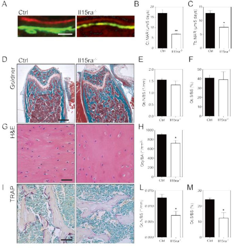
(A) Representative double labeling (scalebar 40μm) of control and il15ra−/− femurs and Mineral Apposition Rate quantification for the cortical (B) and trabecular (C) regions (n=3). (D) Goldner’s trichromic of femur trabecular bone sections (scalebar 500μm) and quantification of osteoblast per bone surface (E) and osteoclast surface per bone surface (F) (n=3). (G) Hematoxylin and eosin staining of femur cortical bone (scalebar 50μm) and quantification of osteocytes/are (H) (n = 3). (I) TRAP staining of femur trabecular bone sections (scalebar 100μm) and quantification of osteoclast per bone surface (L) and osteoclast surface per bone surface (M) (n=3).