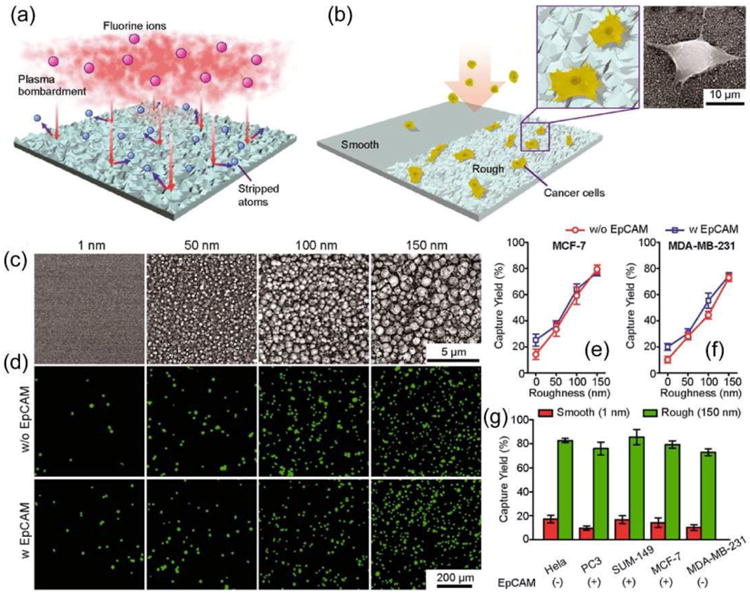
Figure 6.

Intrinsic nanotopological sensing for capturing cancer cells. (a, b) Schematic of nanotopography generated by RIE on glass surfaces. Insets show a zoom-in (left) and SEM (right) images of cancer cells captured on nanorough glass surfaces. (c) SEM images of glass surfaces with their RMS nanoroughness indicated. (d-g) Cancer cells spiked in growth media at a concentration of 105 mL-1 captured on glass surfaces 1 h after cell seeding. Fluorescence images of MCF-7 cells (stained for nuclei, DAPI, green) captured on glass surfaces coated with (bottom) or without (top) EpCAM antibodies (d). Capture yields of MCF-7 (e), MDA-MB-231 (f), and other cancer cell lines (g) as a function of nanoroughness. Reproduced from reference 36 with permission from American Chemical Society, copyright 2013.
