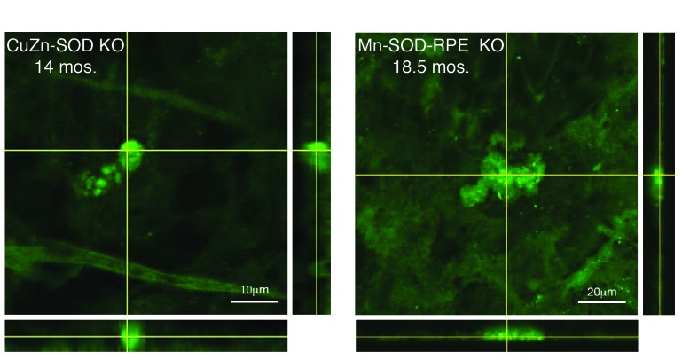
FIGURE 1.
Confocal images of eyecup flat mounts from CuZn-SOD KO and Mn-SOD-RPE KO mice labeled with 10 μM Zinpyr-1. Crosshairs are focused on sub-retinal epithelial drusen-like deposits. The depth of the deposits is visualized by reconstructed optical cross sections (confocal z-stacks) that are displayed along the bottom and right side of each image.

