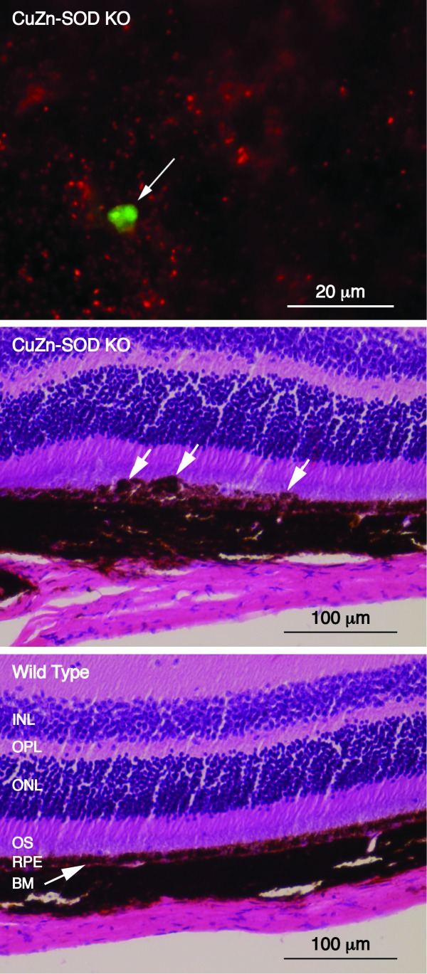
FIGURE 8.
Analysis of CuZn-SOD KO mice. Top; fluorescence micrograph of an eyecup flat mount showing a sub-retinal epithelial deposit stained with ZPP1 (arrow, green) with RPE cell debris (red). Middle; photomicrograph showing the histological analysis of the opposite eye of the same animal. Sub-retinal epithelial deposits are indicated with white arrows. Bottom; histological analysis of an age-matched, wild type control mouse showing normal morphology. INL, inner nuclear layer; OPL, outer plexiform layer; ONL, outer nuclear layer; OS, outer segments of photoreceptor cells; RPE, retinal pigment epithelial cells; BM, Bruch’s membrane.

