Figure 1.

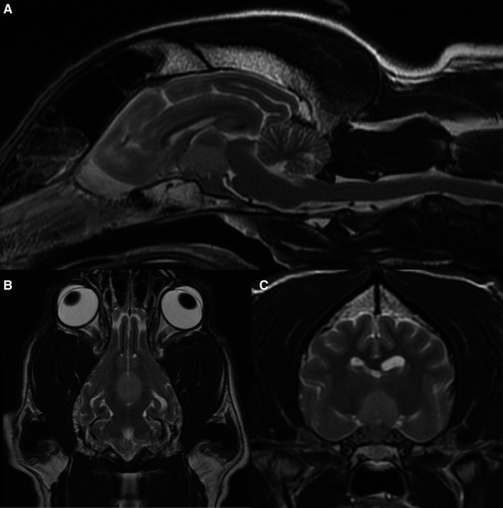
Midsagittal (A), dorsal (B), and transverse (C) T2‐weighted images of the brain. There is a well‐defined, slightly hyperintense to the normal cortical gray matter central mass in the hypophysis/hypothalamic region.

Midsagittal (A), dorsal (B), and transverse (C) T2‐weighted images of the brain. There is a well‐defined, slightly hyperintense to the normal cortical gray matter central mass in the hypophysis/hypothalamic region.