Figure 3.

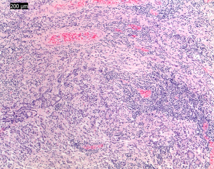
Light micrographs of paraffin‐embedded section stained with hematoxylin and eosin. Diffuse lymphocytic inflammation of the hypophysis. Bar indicates magnification (×10).

Light micrographs of paraffin‐embedded section stained with hematoxylin and eosin. Diffuse lymphocytic inflammation of the hypophysis. Bar indicates magnification (×10).