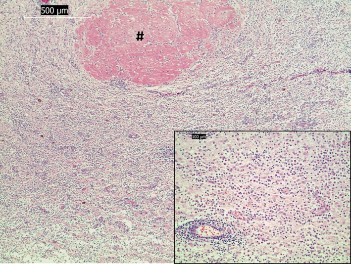
Figure 4.

Light micrographs of paraffin sections stained with hematoxylin and eosin. Diffuse lymphoplasmacytic inflammation of the hypothalamus with perivascular cuffing (# shows the nonaffected left rostral commissure) (×5 magnification). Inset: higher magnification (×20) of infiltrating leukocytic cell population. Bar indicates magnification.
