Figure 3.

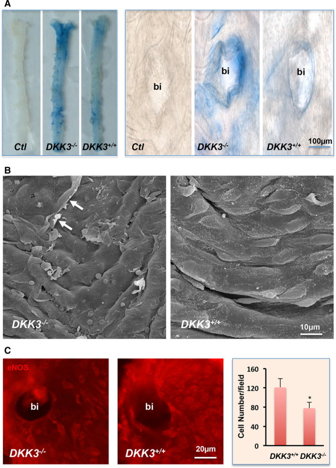
Increased endothelial damage in DKK3-/-/ApoE-/- mice. A, Evans blue dye leaking study. Ten-week-old DKK3-/-/ApoE-/- and DKK3+/+/ApoE-/- mice received an injection of 1% Evans blue dye by the tail vein. Mice were euthanized, and aortas were harvested and washed. Blue areas, representing the damaged area on the surface of aortas were observed. B, Scanning electron microscopy analysis of aortic samples from DKK3-/-/ApoE-/- and DKK3+/+/ApoE-/- mice. Arrows indicate the damaged cell. C, En face preparation of the vessel was stained for endothelial marker eNOS and visualized after incubation with secondary Cy3 conjugated antibody. Quantitative data analysis of the number of endothelial eNOS-positive cells around the bifurcation (bi) areas are presented in the graph (n=6). *P<0.05, indicating significant difference between the 2 groups. ApoE indicates apolipoprotein E; DKK3, dickkopf-related protein 3; and eNOS, endothelial nitric oxide synthase.
